


Author: Vincent Y. Ng, MD
Description
Imaging plays an important role in diagnosing and treating bone and soft-tissue tumors. Imaging studies help physicians determine where a tumor is located, how large it is, and how it affects nearby bones, muscles, nerves, blood vessels, and joints. Certain imaging features may suggest whether a tumor is likely to be slow-growing or more aggressive. When earlier images are available, comparing them can also show whether the tumor has changed over time.
In some cases, imaging strongly suggests a specific diagnosis. However, imaging alone cannot always determine whether a tumor is benign (noncancerous) or malignant (cancerous). Additional evaluation may include blood tests or a biopsy, in which a small sample of tissue is removed and examined under a microscope. When a biopsy is needed, it should be carefully planned—ideally by, or in coordination with, the team that would perform any future tumor surgery. The location and direction of the biopsy can affect the surgical approach and later treatment.
Different imaging tests provide different types of information. Your physician will select the test or combination of tests that is most appropriate for your condition. Imaging studies are usually reviewed by a radiologist, a physician who specializes in interpreting medical images. The radiologist provides a report to the physicians caring for you.
X-rays
Plain radiographs, commonly called X-rays, are often the first imaging study obtained when a bone tumor is suspected. X-rays use a small amount of radiation to create images of the skeleton. Usually, several images are taken from different directions, such as front and side views.
X-rays provide excellent information about bone but less detail about muscles and other soft tissues. They can show how a tumor affects the bone and may provide clues about its behavior. For example, a slow-growing tumor may allow the bone time to form a smooth border, thicken, or remodel around it (like how a tree trunk may remodel around a heavy stone leaning against it). A more aggressive tumor may destroy bone or create a less clearly defined border. Some tumors produce calcium or bone-like material that can also be seen on X-rays.
Fig. 1
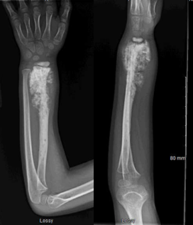
Radiographs, front and side view, of a forearm showing a bone tumor of the distal radius with features concerning for cancerous tumor.
CT scans
Computed tomography, also called CT or CAT scanning, uses X-rays and computer processing to create detailed, cross-sectional images of the body. One way to think about the difference is that a regular X-ray is like a photograph of an entire loaf of bread, while a CT scan shows individual slices of the loaf. These images can also be reconstructed in different directions or as three-dimensional images.
CT scans are particularly useful for showing fine details of bone, including mineralization within a tumor and subtle changes in the bone’s outer surface. They may also be used to evaluate the lungs or other parts of the body when physicians need to determine whether a malignant tumor has spread.
CT scans use more radiation than standard X-rays, but the amount is adjusted to obtain the necessary information while limiting exposure. In some cases, an intravenous, or IV, contrast material is used to make blood vessels and soft tissues easier to see. CT contrast is generally safe, but precautions may be necessary for patients with significant kidney disease or a history of contrast reactions.
MRI scans
Magnetic resonance imaging, or MRI, provides detailed images of muscles, fat, nerves, blood vessels, joints, and bone marrow. It is often the most useful study for determining the full extent of a bone or soft-tissue tumor and its relationship to nearby structures. This information is especially important when planning surgery or other treatment.
MRI does not use ionizing radiation. Instead, it uses a strong magnetic field and radio waves to create images. The examination usually takes longer than an X-ray or CT scan, and the patient must remain still inside the scanner. Some people experience claustrophobia, but strategies such as music, relaxation techniques, medication, or—in selected locations—larger or more open scanners may help.
Because MRI uses a powerful magnet, patients should tell the imaging team about any metal or electronic device in their body. Many joint replacements, surgical implants, and newer pacemakers can be scanned safely, but some devices require special precautions or may not be compatible with MRI. The imaging team will determine whether the examination can be performed safely.
An IV contrast material containing gadolinium may be used to help characterize a tumor. MRI contrast is different from CT contrast and is generally safe. However, the care team may need to take additional precautions in patients with severe kidney disease or a previous contrast reaction.
Fig. 2
MRI of both thighs showing a mass concerning for soft tissue sarcoma on the left thigh.
PET scans
Positron emission tomography, or PET, uses a small amount of a radioactive tracer to show metabolic activity within the body. PET is commonly combined with CT in a single examination, called a PET/CT scan.
The most frequently used tracer is fluorodeoxyglucose, or FDG, which is similar to glucose, a type of sugar. Tissues that use more glucose may take up more FDG. Many cancers show increased FDG uptake because their cells are metabolically active.
The amount of tracer uptake may be reported as a standardized uptake value, or SUV. PET scans can help evaluate the activity and extent of some tumors, identify possible disease in other parts of the body, and assess response to treatments such as chemotherapy or radiation therapy.
However, increased FDG uptake does not always mean cancer. Infection, inflammation, healing tissue, and some benign tumors can also have increased uptake. In addition, some cancers may show little FDG uptake. PET results must therefore be interpreted together with other imaging studies, the patient’s medical history, physical examination, and—when needed—biopsy results.
More Information:
Medscape / eMedicine - Radiology Articles (presentation, history, clinical details, interpretation, imaging, findings, techniques, accuracy, diagnosis, intervention) - Medscape Reference
Radiopaedia - Radiopaedia.org, the peer-reviewed collaborative radiology resource
This is not intended as a substitute for professional medical advice and does not provide advice on treatments or conditions for individual patients. All health and treatment decisions must be made in consultation with your physician(s), utilizing your specific medical information. Inclusion in this is not a recommendation of any product, treatment, physician or hospital.