
Author: Ty Reasnor, MD and Rashmi Agarwal, MD
Description
Synovial chondromatosis is a benign condition that usually affects the joints of middle-aged adults. The disease most commonly is found in the knee joint; however, any joint can be affected. Synovial chondromatosis begins when the inner lining of a joint, called the synovial layer, thickens and creates “balls” or nodules of cartilage. These cartilage nodules can break free and float around as loose bodies in the joint causing joints to “lock up” and cause significant pain.
Symptoms
Patients affected by synovial chondromatosis usually present with pain affecting one joint. The pain can happen at rest and may worsen with movement. In addition to pain, patients can lose flexibility or motion, experience swelling, or feel the joints buckle or give way. Depending on the size and location of the loose body, patients may describe a grinding sensation or feel like something is blocking their knee from moving
Examination
While examining patients with synovial chondromatosis, many of the findings overlap with arthritis. The affected joint may be red, swollen, or tender to the touch. Patients may be able to hear and feel “clicks and pops” in the affected joint. When a doctor is bending or moving the affected joint, he or she may notice that the joint is stiff and not as flexible.
Tests
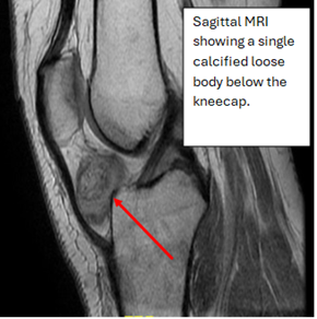
After performing a thorough physical exam, an X-ray of the affected joint is usually obtained. Early on, X-rays usually don’t show the cartilage nodules, but over time, the cartilage hardens or calcifies and becomes visible on X-ray. The x-rays of advanced cases usually show up as one (Figure 1) or multiple (Figure 2) loose bodies in the joint space. If a patient has symptoms, but x-rays do not show anything abnormal, some doctors will choose to obtain a computer tomography (CT) or magnetic resonance imaging (MRI). Both CT and MRI (Figures 3 and 4) can identify synovial chondromatosis and rule out other diseases that have similar symptoms. If a patient chooses surgery for treatment, the surgeon will send the loose bodies that were taken out for further lab testing.
Images
Figure 1: Lateral X-ray of the Right Knee.
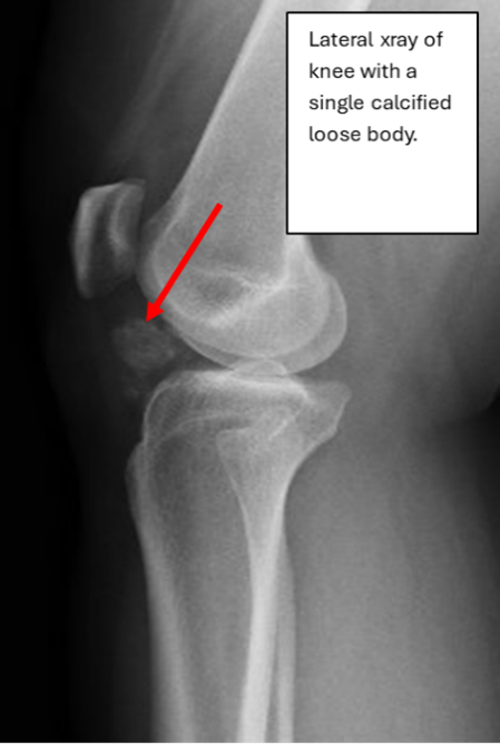
Figure 2: X-ray of a left ankle with multiple loose bodies within the ankle joint.
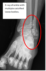
Figure 3: Coronal T1 MRI of a left ankle with multiple loose bodies within the joint.
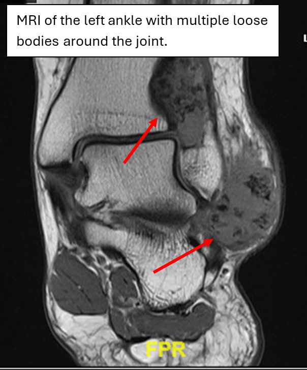
Figure 4: Sagittal MRI of the Right knee.
Prognosis
Generally, the patient’s symptoms and effect on quality-of-life guides treatment. If symptoms are mild, a patient may choose non-invasive or non-surgical treatment. This includes interventions such as ice, heat, anti-inflammatory medications, physical therapy, or injections. If a patient’s symptoms are severe, they may want to proceed with surgery. The surgeon would remove the loose bodies and possibly the inner lining of the joint that is causing the problem. Patients tend to have a significant improvement in symptoms following surgery; however, there is a chance the problem comes back, and patients are seen for follow up over time.
More Information
OrthoInfo from the American Academy of Orthopedic Surgeons:
https://orthoinfo.aaos.org/en/diseases--conditions/synovial-chondromatosis/
Sarcoma Foundation of America:
https://curesarcoma.org/sarcoma-subtypes/synovial-chondromatosis/
This is not intended as a substitute for professional medical advice and does not provide advice on treatments or conditions for individual patients. All health and treatment decisions must be made in consultation with your physician(s), utilizing your specific medical information. Inclusion in this is not a recommendation of any product, treatment, physician or hospital.