Author: Lisa Ercolano, MD
Description
Glomus tumor is a benign tumor made up of cells that resemble smooth muscle cells of the glomus body, which is a thermoregulatory structure of the skin. They are most commonly found in fingers, under the nail, on the fingertip or in the foot, but can occur in any location. These are very rare tumors, accounting for less than 2% of all soft tissue tumors. Most of these tumors occur before the age of 30, though very rarely in children. More rarely, there are cases of multiple congenital glomus tumors which are often symptom free but associated with other syndromes or findings.
Symptoms
The most common symptom is intermittent pain out of proportion to the size of the mass. Patients can present with small nodules under the skin which may vary from red to purple to blue depending on how deep they are. Often however, the mass is not obvious, and the pain source remains unclear. Pressure can worsen symptoms. If exposed to cold, they may trigger a temperature change of the entire extremity as well. These symptoms can have significant effect on quality of life, especially when prolonged due to missed diagnosis.
Examination
Palpation of a small red/blue/purple mass that reproduces pain, most commonly in the finger should suggest the possibility of a glomus tumor. If that mass is also sensitive to cold temperature, a glomus tumor diagnosis is supported. If the mass is located under the nail, changes in that nail are sometimes present. Occasionally, a mass cannot be palpated, or the tissue is simply thickened while these other symptoms persist. If the pain and tenderness is reduced by inflating a tourniquet to the finger, this also supports the diagnosis.
Tests
X-rays of the affected area are the first step in diagnosis, which can sometimes show bony erosions if the lesion is next to the bone. A CT scan may show these bony changes in more detail and also identify a mass in the soft tissue. Ultrasound can also be used to identify a solid mass. MRI is the best study to identify the mass and define its features. These imaging studies cannot ensure a diagnosis however, and obtaining a biopsy or sample of the mass is the only way to confirm diagnosis.
Images
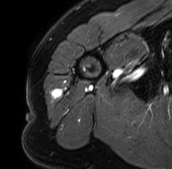
Figure 1. Glomus tumor in the deltoid muscle of the shoulder. It measures less than a centimeter and is otherwise a homogenous solid bright mass in the otherwise normal muscle tissue.

Prognosis
Almost all glomus tumors are benign and are therefore not a cancer concern. The severe and sometimes debilitating pain is cured by surgically removing the mass. There is a recurrence risk of approximately 10% after removal. Alternatives to surgery include sclerotherapy or laser therapy.
More Information:
Glomus Tumor - Pathology - Orthobullets
This is not intended as a substitute for professional medical advice and does not provide advice on treatments or conditions for individual patients. All health and treatment decisions must be made in consultation with your physician(s), utilizing your specific medical information. Inclusion in this is not a recommendation of any product, treatment, physician or hospital.


